Case Report 
 Creative Commons, CC-BY
Intrahepatic Gallbladder Associated with Fitz-Hugh- Curtis Syndrome: A Case Report
*Corresponding author:Hernandez Faruk, General Physician Universidad del Norte Barranquilla, General Surgeon, Universidad Metropolitana de Barranquilla, Fellow of Gastroenterology, Universidad de Cartagena, Colombia.
Received:January 06, 2023; Published:February 15, 2023
DOI: 10.34297/AJBSR.2023.18.002426
Abstract
The intrahepatic vesicle is the most frequent presentation of difficult cholecystectomies. FitzHugh-Curtis syndrome is a discovery, whose incidence tends to increase with the use of laparoscopy; this is in the order of 15-30% in women with pelvic inflammatory disease. This article presents the case of a 34-year-old woman, who has a clinical picture with more than a 30-day evolution, which was characterized by right hypochondriac pain of moderate-intensity, irradiated to the ipsilateral scapular region. The diagnosis was performed through abdominal ultrasonography of multiple cholelithiasis; for this reason, it is decided to perform an elective laparoscopic cholecystectomy, in which was found an intrahepatic vesicle with multiple calculations and besides, adhesions in violin strings between the hepatic capsule and the abdominal wall, concluding a post-surgical diagnosis of FitzHugh-Curtis syndrome.
Keywords: Intrahepatic vesicle, Fitz Hugh Curtis syndrome, Violin string adhesions
Introduction
Intrahepatic gallbladder is one of the most frequent congenital anomalies of ectopic locations, with an incidence of 0.1-0.7% [1]; it is the most frequent presentation of difficult cholecystectomies and in our environment, it is an intraoperative finding since patients with biliary pathologies are taken to surgery only with ultrasound as a diagnostic aid. The surgical approach to an intrahepatic gallbladder is considered a difficult cholecystectomy, that is, a condition in which there are factors associated with the organ, adjacent organs or inherent to the patient that do not allow a fast and safe dissection, which implies a prolongation of the surgical time [2] and in our case a higher risk of intraoperative bleeding.
Currently, Fitz-Hugh-Curtis syndrome (FHC) is a finding whose incidence tends to increase with the use of laparoscopy; no exact statistics are known for cholecystectomies; however, the incidence in laparoscopic surgeries in gynecological patients is in the order of 15-30% and the overall incidence is 4-14% [3]. FHC syndrome, or perihepatitis associated with pelvic inflammatory disease, is a process affecting the hepatic capsule and abdominal wall characterized by the presence of “violin-string” adhesions first described by Cario Stajano in 1920 [4] although it is named after Fitz Hugh and Artur Curtis who described its acute and chronic manifestations [5].
Initially it was related to gonococcal infections by Neisseria gonorrhoeae, but in 1978 Müller Shoop, et al. described Chlamydia trachomatis as the microorganism involved in its pathophysiology [6] and it is currently considered by many authors as the main etiological agent [7]. This entity may present asymptomatically in many cases or with pain and hyperalgesia in the right upper quadrant caused by irritation of Glisson’s capsule in the liver. The objective of this review is to present a case of an incidental finding of chronic stage FHC in a patient with a diagnosis of cholelithiasis whose finding was demonstrated in a laparoscopic cholecystectomy procedure during which an intrahepatic gallbladder was also evidenced.
Presentation of the Case
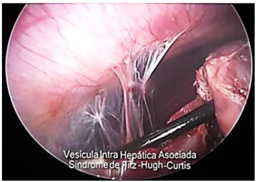
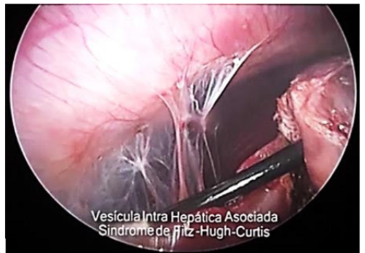
Female patient, 34years old, resident of the city of Sincelejo- Sucre, who works as a companion, with a history of three pregnancies, two childbirths, one abortion and one Pomeroy surgery 3years ago. She was admitted with pain in the right hypochondrium of more than 30days of evolution of moderate intensity and radiating to the right scapula, concomitant with dizziness. Physical examination showed afebrile, soft abdomen, not painful on palpation, positive peristalsis, negative Murphy, and no signs of peritoneal irritation. The ultrasound report showed multiple cholelithiasis and she was scheduled for elective laparoscopic surgery. During surgery, in addition to multiple cholelithiasis, an intrahepatic gallbladder was found, together with the diagnosis of Fitz Hugs Curtis Syndrome proved by direct visualization of the presence of perihepatic adhesions like violin strings (Figures 1,2).

Figure 2:Visualization of the body and fundus portion of the gallbladder within the liver parenchyma.
Due to the risk of fetal abnormalities [9] and chances of miscarriages during invasive prenatal sampling, the use of cell free fetal DNA is considered a more attractive alternative of prenatal diagnosis. Cff DNA is apoptotic fetal DNA that shed from villous trophoblast during the development of fetus [11]. In addition to the role of cff DNA in detection of chromosomal abnormalties and single gene disorders, studies have shown the association of significant increase in the level of cff DNA in maternal plasma during pathological conditions for example scleroderma, [12] preeclampsia, [13,14] trisomy 21 [15] intrauterine growth restriction [16,17] and preterm labor [18]. Hence cffDNA can be used as an alternative screening marker for the detection of pregnancy loss disorders. The objective of this study is to investigate correlation of the concentration of cff DNA with intrauterine deaths
Discussion
The finding of HCFS was not common during surgeries, but with the advent of laparoscopic surgical techniques it has increased, an exact statistic in cholecystectomies is not known, however, the incidence in laparoscopic surgeries in gynecological patients is in the order of 15-30% [3].
In the case presented, it was an incidental finding because the approach was directed to treat a cholelithiasis because the symptomatology referred by the patient was characterized by a colicky pain, located in the right hypochondrium, of 30days of evolution, with a negative Murphy, which radiated to the ipsilateral scapula, It was not suspected that the patient could have HCFS since she did not report having previously presented an inflammatory pelvic infection and because in our environment patients with cholelithiasis are taken to surgery only with ultrasound, which is the method recommended by the Colombian guidelines as the method of choice to make the diagnosis, Therefore, more specialized studies were not performed, which could suggest the presence of HCFS, such as the CT scan that evidences a reinforcement in the hepatic capsule in these patients [8]. The diagnosis of HCFS in our case was by direct visualization of the adhesions in violin strings on the hepatic surface and the abdominal wall during laparoscopic cholecystectomy, which is a pathognomonic sign that occurs in the chronic stage of this entity [7]. There is no clear theory as to how microorganisms ascend from the genital tract to the hepatic capsule, multiple hypotheses have been mentioned, but all of them show inconsistencies, however, the most accepted of them proposes that adhesions are the result of inflammation produced by bacteria that arrive by dissemination from an inflammatory pelvic disease, using the circulation of the peritoneal fluid as the main route until reaching the subphrenic space [7].
The first corresponds to the acute phase in which there is sudden, intense pain at the level of the right costal border that is exacerbated by movement, deep inspiration, and cough, in addition to classic manifestations of PID such as leukorrhea [7]. In the chronic stage, it is characterized by pain and hypersensitivity in the right hypochondrium caused by irritation of Glisson’s capsule [3].
Regarding the association of HCFS with cholelithiasis, this is based solely on the fact that both present with pain in the right hypochondrium and can be referred to the ipsilateral shoulder, it is important to note that there is no pathological association, however, making the differential diagnosis is important because there are cases that can be framed as happened in our patient, so it is important to make a good anamnesis and do not forget to ask about history of pelvic diseases or sexually transmitted infections.
Another incidental finding found in the patient was an intrahepatic gallbladder, a congenital disorder that occurs at the stage of gallbladder formation; Normally this is formed within the hepatic parenchyma in the fetal stage, and by the second week of gestation it takes its definitive position outside the hepatic parenchyma [9] , in the gallbladder bed, when this does not happen what is known as intrahepatic gallbladder occurs, this disorder has an incidence of 0.1-0.7% [1] and is associated with an altered function of the organ that predisposes to the formation of gallstones by biliary stasis.
The patient underwent cholecystectomy by laparoscopy and adhesiolysis was performed so that the pain would not continue, since it is not possible to know which of the two pathologies triggered the pain in the right hypochondrium because both manifest themselves in the same way or if the pain was caused by both pathologies at the same time. Adhesiolysis is not indicated in all patients, only in those in which the clinical picture does not yield with medical management. In the case of the patient, as it was an incidental finding which was not suspected at the beginning, the surgical procedure was performed and then she was treated with antibiotic doxycycline caps 100mg VO for 14days and ceftriaxone 500mg IM single dose, it is also recommended to treat the sexual partner in all cases to avoid recurrences [7].
References
- Pérez OJ, Haito Ch Y, Rodríguez VF (2015) Intrahepatic Gallbladder: Intraparenchymal Approach. Chilean Journal of Surgery 67(4).
- Alvares FL, Rivera D, Esmeral EM, Garcia CM, Toro FD, et al. (2013) Difficult Laparoscopic Cholecystectomy, Management Strategies. Colombian Journal of Surgery 28(3).
- Lozano DM, Jiménez HJ, Hernández GR (2009) Prevalence of Fitz-Hugh-Curtis Syndrome by Laparoscopy in Patients of The Gynecology Service at The Hospital Juarez De Mexico. Journal of the Hospital Juarez De Mexico 76(1): 23-27.
- Ricci AP, Solà DV, Pardo SJ (2009) Fitz-Hugh-Curtis Syndrome as A Finding During Gynecologic Surgery. Chilean Journal of Obstetrics and Gynecology 74(3).
- Bearse C (1931) Ghonorrheal Salpingitis and Perihepatic Adhesions. Jama 97(19).
- Müller Schoop JW, Wang SP, Munzinger J, Schläpfer HU, Knoblauch M, et al. (1978) Chlamydia Trachomatis as Possible Cause of Peritonitis and Perihepatitis in Young Women. British Medical Journal 1(6119).
- Ramirez CG, De La Peña MS, Ramirez AC, Antonio Liho NA (2009) Fitz-Hugh-Curtis Syndrome. A Case Report and Review of Literature. Endoscopic Surgery 10(3,4).
- Nishie A, Yoshimitsu K, Irie H, Yoshitake T, Aibe H, et al. (2003) Fitz-Hugh-Curtis Syndrome. Journal Of Computer Assisted Tomography 27(5).
- Audi P, Noronha F, Rodrigues J (2009) Intrahepatic Gallbladder A Case Report and Review of Literature. The Internet Journal of Surgery 24(1).


 We use cookies to ensure you get the best experience on our website.